
A new find of sapphire in Ranotsara, in the south of Madagascar, reportedly unheated after the miner. The gemstone, figure 1, is bluish-purple while viewed down to the table but pinkish-purple other orthogonal directions. The color is also subject to change depending on the light source being used to view the stone. The color shift is bluish-purple in cool white (day light) although it is strong purple in warm white.
The gemstone has some minute inclusions that can be observed in figure 1.
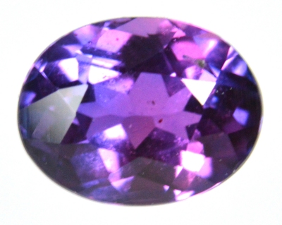 Figure 1. New sapphire from Ranotsara, Madagascar - 0.49 ct.
| Shape | oval |
| Size | 5.0 x 3.8 x 2.8 |
| Color | bluish-purple |
| Lustre | bright-vitreous |
| Weight | 0.49 |
| SG | - [3.95 - 4.05] |
| RI | 1.762 - 1.770 [1.760 - 1.775] |
| DR | 0.008 U- [0.008-0.009 U-] |
| Pleochroism | bluish-purple / light-pinkish-purple |
| Polariscope / Conoscope | C-axis perpendicular to the table, interferences figures are visible without conoscope. Uniaxial |
| SWUV | inert |
| LWUV | red |
| Magnetic susceptibility | inert |
Table 1. Observational and measured properties (typical values for the specie are enclosed in square brackets [...])
Infrared reflectance spectroscopy:
The IR reflectance spectrum shown in figure 2 was acquired from the gemstone table. The spectrum pattern is typical for corundum (ruby and sapphire).
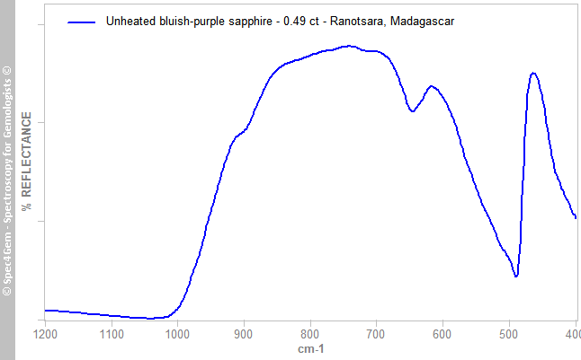 Figure 2. IR reflectance spectrum of the Ranotsara unheated bluish-purple sapphire acquired from the table which is characteristic of corundum.
UV-VIS-NIR spectroscopy:
The UV-Vis-NIR absorption spectrum, figure 3, was acquired with a light path entering the pavilion in direction of the table, thus the light path is almost parallel to the C-axis.
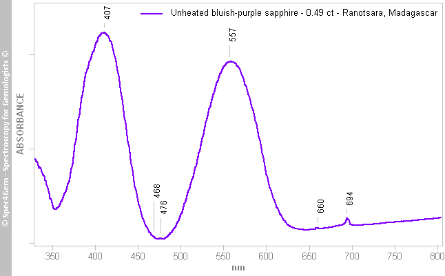 Figure 3. UV-Vis-NIR spectrum of the unheated bluish-purple sapphire from Ranotsara, Madagascar. The pattern is typically that of Cr3+ with two 407 and 557 nm strong absorption bands, and the additional weaker bands at 468, 476, 680 and 694 nm.
 The spectrum of figure 3 shows the cause of color is Cr3+ with its two 407 and 557 nm strong absorption bands and the additional weaker bands at 468, 476, 680 and 694 nm. Interestingly, within the transmission window created by the two strong Cr3+ bands, the absorbance is lower (~ the transmittance is higher) than the absorbance (respectively the transmittance) measured in the orange and red spectral region.
The sharp spin-forbidden 694 nm band appears like an absorption band and not like an emission band as often seen in nowadays spectrum. If the spectrum is acquired with a CCD spectrometer, all the white light spectrum is used at a time and its violet component can excite the red luminescence of Cr3+ causing an emission band thus a negative absorption band. If a monochromator is used to get the spectrum, the stone gets the light for a given wavelength at a time, so while measuring the absorption at 694 nm, the stone is not excited by the violet light and there is no induced luminescence with an emission of light resulting in a negative band.
There is no Fe3+ feature visible in the spectrum, this does mean there is no Fe3+ in the stone but if it exists its features are hidden by those of Cr3+.
Another question can be raised about V3+ (trivalent vanadium) that might exists in corundum. If the Cr3+ absorption bands are generally observed at 405-410 and 550-560 nm, those of V3+ are at 400 and 575 nm. The figure 3 spectrum shows well defined band for Cr3+ even if weak shoulders could exist around 380 and 680 nm. A simulation of the possible bands was processed by deconvolution but the result is not convincing since the band fitting gives bands at 387, 417, 542 and 575 nm. Cr3+ bands are thus shifted to 417 and 542 nm that is highly unlikely. Another guess gives bands at 387, 415, 555 and 583 nm, that is a bit better for the Cr3+ bands at 415 and 555 nm with V3+ bands being 387 and 583 nm. These values are a bit far from the published data for V3+. The UV-Vis spectrum does not gives any evidence of the V3+ presence in the structure, it might be there but it does not significantly affect the stone's color mainly resulting of Cr3+ presence.
One can ask why the stone is not red like a ruby since it has a Cr3+ spectrum? Within the ruby the transmission window around 475 nm is not as deep as it is for this sapphire. As a consequence, this sapphire material transmit more blue than a ruby, creating the bluish-purple color. The blue transmission could be partially removed by heating.
Photoluminescence spectroscopy:
The photoluminescnce spectrum, figure 4, was acquired with a 405 nm laser source having its beam path oriented perpendicularly to the C-axis and parallel to length of the stone.
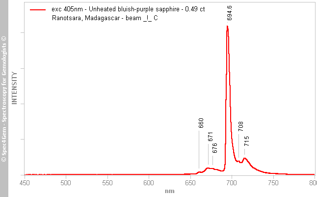 Figure 4. Photoluminescence spectrum of the unheated bluish-purple sapphire excited by a 405 nm laser with the laser beam path perpendicular to the C-axis. The result is the well known Cr3+ emission spectrum with the sharp 694.6 nm emission peak.
The 405 nm excitation source does not induce any luminescence except that of the typical Cr3+ with its strong and sharp peak at 694.6 nm along its much weaker side peaks at 680, 671, 676, 708 and 715 nm.
Conclusion:
This bluish-purple sapphire from Ranotsara, south of Madagascar has common properties except it owes its color to Cr3+ like rubies but with an additional blue component. The UV-Vis-NIR absorption spectrum does not reveal any evidence of Fe3+ iron common in sapphire.