
Since 2015 spring, there has been some rumor about red kornerupine coming from Madagascar. Quickly, the material was identified as cordierite being the first occurrence of red cordierite. The material comes from the Iakora district, Fianarantsoa province, in southeast Madagascar. The figure 1 shows the sample of this particular gemstone, faceted for a total weight of 2.96 ct. The color is hard to define because it varies a lot depending on the direction it is observed from. Overall color is brownish-orange but deep orange-red can be observed as well as light brownish-yellow.
According to some other information sources, brown and orangy-brown cordierite was already but seldom mined a decade or two ago, unfortunately there is few documentation about it. The material was mainly for collectors, a sample was described in the Winter 2004 issue of Gems & Gemology[1].
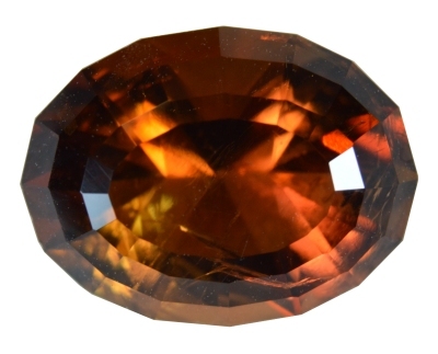 Figure 1. 2.96 ct brownish-orange cordierite from Iakora, Madagascar
| Shape | oval step cut |
| Size |
11.3 x 8.6 x 6.4 mm |
| Color | brownish-orange |
| Lustre | vitreous |
| Weight | 2.96 ct |
| SG | 2.55 — [2.52 - 2.79] |
| RI | nα=1.529, nβ=1.533/1.534, nγ=1.538 — [1.520 - 1.580] |
| DR | 0.009 B +/- — [0.005 - 0.018, B-, sometime B+] |
| Pleochroism | orange-red/light brown-yellow, orange-red/violetish-blue, light brown-yellow/violetish-blue |
| Polariscope / Conoscope |
biaxial |
| SWUV | very faint red |
| LWUV | inert |
| Magnetic susceptibility | inert |
Table 1. Observational and measured properties (typical values for the specie are enclosed in square brackets — [...])
Cordierite is famous for its strong trichroism especially the purplish-blue one known as iolite. As expected, as for other cordierite, this cut gemstone shows a pleochroism even without polarizing filters. Using a london dichroscope helps in getting the true colors even if it is not that easy with such a cut stone. Because of the cut which has lot of marked angles, the observation of light in a given direction is sometime tricky. Another important property is that the color saturation strongly increase with the thickness of the material. Getting a piece of rough would be a better solution to observe the pleochroism.
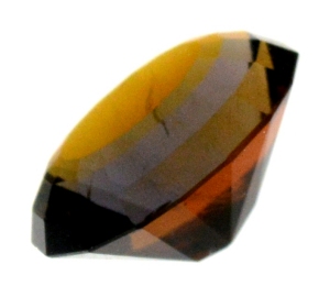 Figure 2. All three colors together through the a
polarizer, color mixing explains the medium
saturation / hue.
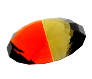 Figure 3. The strong orange-red and the light
brownish-yellow colors together through london
dichoscope.
The figure 2 shows the stone through a single polarizer, three colors are observed, orange, purplish-blue and yellow. The colors as they are not fully separated - distinct from each other - they mix together and the result is a medium saturation : hue. So instead of viewing the deep orange-red, it is a brownish-orange (unsaturated orange), instead of light brownish-yellow it is a medium orange-yellow, the purplish blue does not really differ from the fully saturated one.
The figure 3 shows an example of a color couple through the polarizers filters of a London dischroscope, the deep orange-red and the light brownish-yellow. Not all color couples have been photographed.
All optical an physical properties, table 1, fall in the known ranges for cordierite. An interesting feature is the optical character sign is undefined or positive/negative, in other words nβ=1.533/1.534 is just in the middle of nα=1.529 and nγ=1.538, a reading error as low as 0.001 can change the sign in this particular case, so this sign measurement is inconclusive. Usually, the cordierite is biaxial negative but regarding this new material, other publications relate the uncommon positive optical character sign, with the following values: nα=1.530, nβ=1.533/1.534, nγ=1.540[1] and nα=1.528/1.530, nβ=1.532/1.533, nγ=1.538/1.540[2].
Infrared reflectance spectroscopy:
Two infrared reflectance spectra were acquired, the first one from the table and the last one from a pavilion's facet nearly perpendicular to one of the optical axis. Both spectra are presented in figure 4, few differences are observed. The spectra are consistent with other cordierites' spectra, characteristic of the mineral specie.
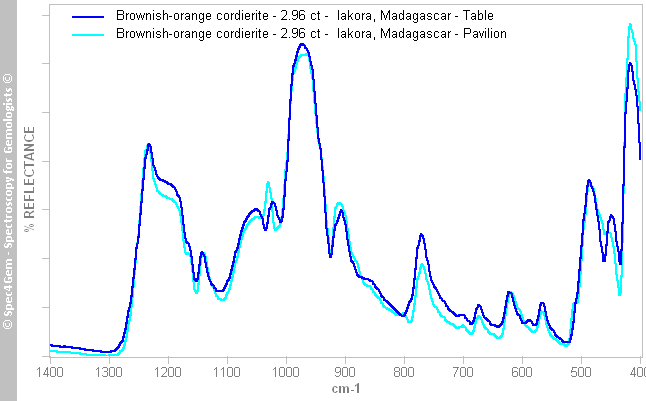 Figure 4. IR reflectance spectra acquired from the table and from a pavilion's facet nearly perpendicular to one of the optical axis. The spectra are characteristic of the cordierite mineral specie.
UV-VIS-NIR spectroscopy:
A first UV-Vis-NIR spectrum, figure 5, was acquired with a light path crossing the stone from the culet to the table. This direction does not correspond with an optical axis. A large transmission window almost centered on 700 nm is formed by a continuum absorption towards the UV with a large absorption band around 510-520 nm and a strong absorption band in the NIR centered around 950-980 nm. Such a spectrum produces the brownish-orange/red color by mostly removing the violet, blue and partly the green and yellow, letting the orange and red transmitted.
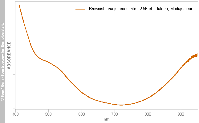 Figure 5. The UV-Vis-NIR spectrum acquired with a light path crossing the stone from the culet to the table responsible for the brownish-orange color because of the large transmission window centered on 700nm.
The cordierite being trichroic, it can be interesting to compare the spectra obtained for the three colors at their maximum when light is polarized parallel with a crystallographic axis. This can easily be achieved when the sample is a rough that can be sawn / sliced according to the crystallographic axis. With the cut stones, except in rare cases, the easiest solution is to use polarizing filters as this is done while looking for pleochroism's colors with a dichrocope.
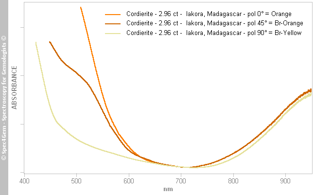 Figure 6. UV-Vis-NIR spectra obtained with a polarizer oriented to get the orange (0°) and the light brownish-yellow colors (90°) maxima. An intermediate spectrum was acquired with the polarizer rotated of 45°, the spectra is similar to that of figure 5 acquired without the polarizer.
 Figure 6, three spectra were acquired with the same light path used as for that of figure 5, but the first one with the polarizer oriented to get the maximum of the orange/red color, the second one with the polarizer rotated of 45° and the last one with the polarizer rotated of 90° to let the light brownish-yellow color transmitted. The absorbance varies significantly between 450 and 600 nm from a weak absorption of the blue to the yellow to a full absorption (cut-off) of the blue to the green-yellow. This differential absorption explains the strong pleochroism from the brownish-yellow to brownish-orange/red.
Observing the variation of the spectra in the following order: 'pol 90°' → 'pol 45°' → 'pol 0°', a centered band around 520 nm rises, creating the cut-off. This band is responsible for the strong pleochroism.
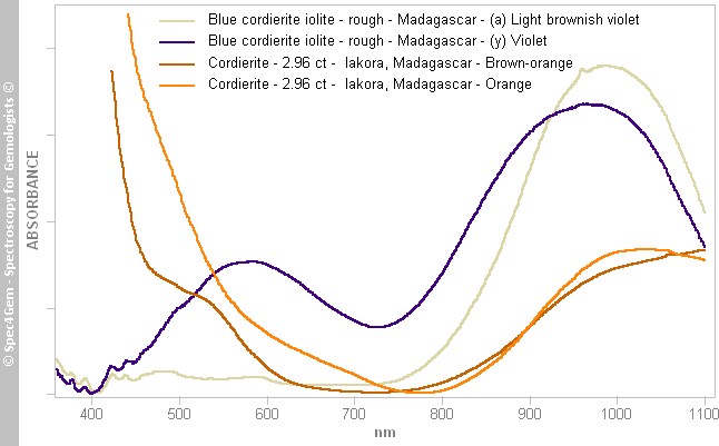 Figure 7. The UV-Vis-NIR spectra acquired for the orange and brownish-orange light path of the brownish-orange cordierite without polarizing filter but up to 1100 nm. They can be compared to the spectra of the α and γ direction of a violet/blue iolite from Madagascar. Spectra pattern are fully different on the 350 to 750 nm but almost similar on the NIR range.
The spectrum pattern is far different from the spectrum of the violet/blue cordierite known as iolite as shown in figure 7. The iolite spectra have weak absorptions in the ultra-violet to the green region (370 nm - 500 nm) ascribed to the spin forbidden transition of Fe2+, Fe3+, Mn2+ and Mn3+[4] which are almost independent of light polarization. A large band centered around 590 nm – called the pleochroic band – appears at its maximum in the γ spectrum (light beam || a crystal-axis) and partially in the β spectrum (light beam || b crystal-axis) but the later is inexistant in the α spectrum (light beam || c crystal-axis). The only part of the spectrum which is similar to both cordierite, the brownish-orange and the violet/blue ones, is the growing absorbance towards the NIR starting at 750 nm which usually correspond to a strong band centered between 965 and 995 nm. However, the later band of the brownish-orange cordierite does not have the maximum there. Either it is around 1030 nm or further because absorbance does not seem to decrease towards longer wavelength. It is not clear whether the slope down does not really exist or it is a wrong measurement due to the spectrometer's limits.
Even if the acquired spectra are rather significant, getting better polarized spectra would require a rough sample of this cordierite material to slice / cut it according to the crystallographic axis.
Photoluminescence spectroscopy:
A red luminescence can be observed with a 20 mW 405 nm laser but it is very weak and localized on the laser beam path in the stone as shown in figure 8. The same luminescence can be observed with a 1 mW 411 nm laser, still weak but not as weak as with the 405 nm. The 411 nm wavelength is likely closer to an excitation feature than the 405 nm one. The emission spectrum is shown in figure 12 (light red spectrum). The luminescence also depends on the polarization.
The SWUV lamp (254 nm) gives a faint red luminescence as shown in figure 11, even if in a first time the doubt is possible because such a lamp even equipped with a filter lets some peaks transmitted in the red and deep red spectral region. Therefore, when the red color is observed it is never clear if it is the result of luminescence or the light emitted from the SWUV lamp. Observing the deep red light with the spectrometer gives the answer because the deep red light is the result of an asymmetric bell shape emission (a gaussian) with the added SWUV red peaks (sharp peaks). Both deep red light cause can be then distinguished. The emission spectrum shown in figure 12 (dark red spectrum) was processed to clean / remove the spurious red peaks of the SWUV lamp.
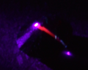 Figure 8. Red luminescence on the 405 nm laser
beam path.
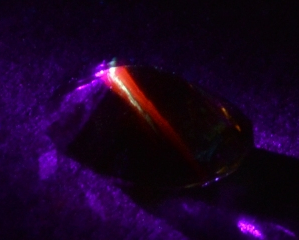 Figure 9. Red luminescence on the 405 nm laser
beam path surrounding the white luminescence of
a healed crack.
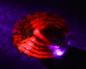 Figure 10. Red luminescence on the 405 nm laser
beam path reflected in the gemstone (viewed table
up). The laser beam path is seen in the middle.
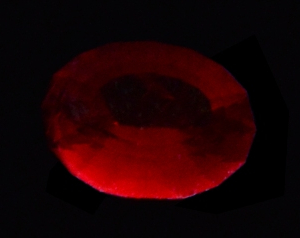 Figure 11. Red luminescence induced by the
254 nm SWUV lamp (viewed table up).
Other excitation sources were experimented without success, all of them have a weak power compared to the laser and SWUV lamp, a 2 mW 280 nm source, a 370 nm source and a 1 mW 532 nm laser source. Either the emission wavelength of these sources do not match the excitation wavelength of the material or they are not powerful enough to bring the required energy to get the material excited. The 532 nm source produces an emission around 646 nm ascribed to Mn2+ and sometime a sharp peak at 658 nm ascribed to Cr3+[6]. Other sources especially in the violet, violet-blue should be tried, for example around the 445-450 nm where the Mn2+ and Mn3+ absorption are usually stronger. Although a 632-635 nm source was used for the violet/blue cordierite[4] because it matches the 'pleochroic band' and it produces the same kind of emission band at 690 nm, such source was not tried, because the wavelength correspond to the minima of absorption of this brownish-orange cordierite, so it should not match any excitation wavelength.
Here again, an easier way to get more precise results would required a piece of rough to be able to observe the luminescence depending on the polarization.
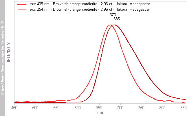 Figure 12. Photoluminescence spectra of the 2.96 ct brownish-orange cordierite from Madagascar excited by a 405 nm laser (light red spectrum) showing an emission peak at 676 nm and excited by a 254 nm SWUV lamp showing an asymmetric emission peak at 685 nm. Due to the very weak luminescence, acquiring the spectra was somewhat difficult.
The 405 nm excited luminescence is a quite large emission viewed as red which is composed of a single emission peak at 676 nm, a gaussian fitting gives 678 nm.
The 254 nm excited luminescence is an asymmetric emission peak at 685 with the wing toward the NIR.
If we consider the 254 nm luminescence spectrum as the result of the sum of at least two gaussian emission band (this is likely incorrect but for simplification), a band fitting gives two bands at 678 and 723 nm. Each excitation source, with the later assumption for the 254 nm excitation, seems to induce the same luminescence emission at 678 nm.
The 678 nm emission could be ascribed to Mn3+ as described by Spinolo et al[4] for the violet/blue cordierite. The possible but not proved emission at 723 nm does find any explanation, Fe3+ is expected at 750 nm[5].
In figure 9, a quite strong yellow luminescence excited by the 405 nm laser was also observed along the healed crack, the luminescence spectrum is available in figure 13. The yellow luminescence observed in the crack is surrounded by the red one because of the limits of the spectrometer, the laser beam cannot be focused only on the crack and the detector captures an area larger than the crack, detailed analysis would require a confocal microscope to focus and and analyze the same focal plane. It is important to understand that the cracks are within the stone, they do not crop out the surface, so the laser beam has to cross the gemstone material before reaching the crack, that also explains the surrounding red luminescence.
The acquired spectrum, figure13 (yellow spectrum), shows two emission peaks at 590 and 666 nm, the 590 nm emission causing the observed yellow color and the 666 nm peak is the already observed red luminescence which is inherent to this cordierite material. A peak fitting gives the peaks at 588 and 679 nm, confirming the 666 nm peak as being definitely the 678 nm one. The cause of the yellow luminescence has not been investigated yet.
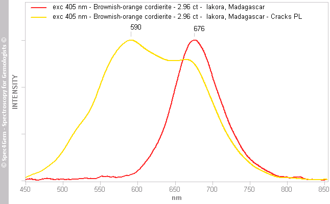 Figure 13. Photoluminescence spectra of a crack within the 2.96 ct brownish-orange cordierite from Madagascar excited by a 405 nm laser (yellow spectrum) showing an emission peak at 590 and 666 nm. The red luminescence spectrum (light red spectrum) is displayed for comparison.
Conclusion:
The results of the classical gemology are consistent with the published one by the different authors, except the red color that cannot be observed without a dichroscope and one of the pleochroic colors which is light violetish-blue instead of being almost black as described by Fritsch et al[3]. The cordierite mineral specie was confirmed by the infrared reflectance spectroscopy.
The pleochroic (polarized) UV-Vis-NIR are different of those observed in the violet/blue cordierite namely iolite. The UV and violet is always absorbed and a strong dichroic band rising at 520 nm create the 600 nm cut-off resulting in the orange-red color. Color causes are not identified yet.
The photoluminescence differs of that usually observed except that described by Spinolo et al[4] for the violet/blue cordierite which is ascribed to Mn3+ with an emission at 689 nm, in this brownish-orange cordierite it is at 678 nm, with only 10 nm far away. Mn3+ is possibly the cause of the red luminescence. The yellow luminescence at 589 nm from the cracks/inclusions has not been investigated yet. The 723 nm luminescence is not explained if this later is proven to exist and is not an artifact caused by the method used with the SWUV lamp.
To go further, a rough piece of this cordierite material is required.
[1] Orangy Brown IOLITE From Madagascar, Elizabeth P. Quinn, Gems & Gemmology, 2004, Winter, p. 328
[2] Orange-Red to Yellowish Brown Cordierite from Madagascar, J. C. (Hanco) Zwaan, Journal of Gemmology, 2016, volume 35, No.1, pp. 8–9
[3] Red cordierite from Madagascar, E. Fritsch, B. Rondeau, T. Peclet, P. Lefebvre, Y. Lulzac, Gems & Gemmology, 2016, Spring, pp. 97–98
[4] Mn sites in cordierite - electron paramagnetic resonance, luminescence, and optical absorption analysis, G Spinolo, S. Gabrielli, V. Palanza, R. Lorenzi, M.C. Mozzati, M. Fasoli, N. Chiodin, B. Vodopivec and A. Paleari, Eur. J. Mineral. 2012, 24, 447–456
[5] Luminescent Spectra of Minerals, Boris S. Gorobets and Alexandre A. Rogojine, Moscow, 2002, ISBN: 5901837053
[6] Modern Luminescence Spectroscopy of Minerals and Materials, 2nd Edition, M. Gaft, R. Reisfeld, G. Panczer, Springer Editor, ISBN: 9783319247632